- Hemocytometer
-
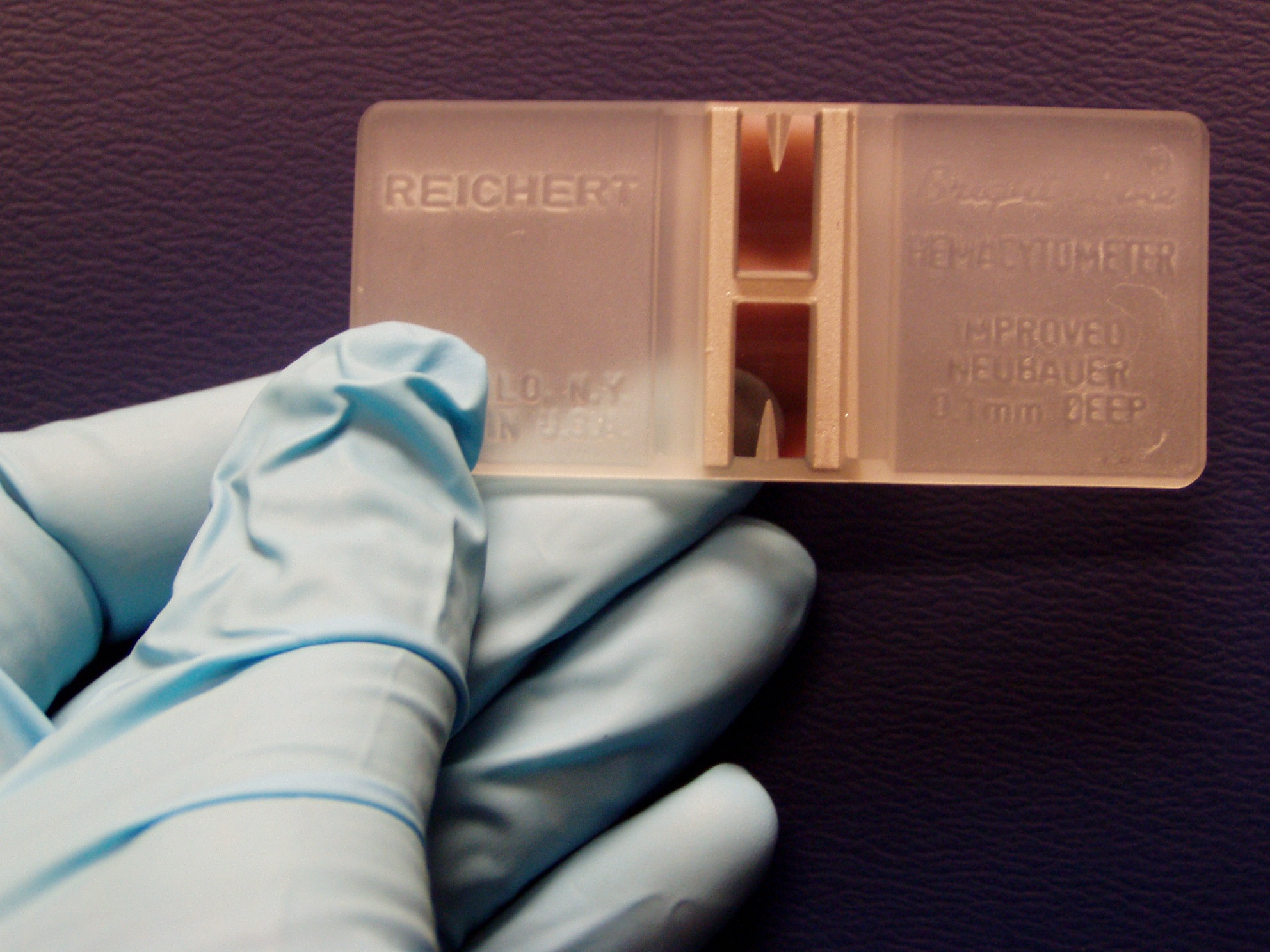
 A hemocytometer. The two semi-reflective rectangles are the counting chambers.
 Load a chamber
 The parts of the hemocytometer (as viewed from the side) are identified.
 Hemocytometer grid:
red square = 1 mm2, 100 nl
green square = 0.0625 mm2, 6.25 nl
yellow square = 0.04 mm2, 4 nl
blue square = 0.0025 mm2, 0.25 nl
at a depth of 0.1 mm.The hemocytometer or haemocytometer is a device originally designed for the counting of blood cells. It is now also used to count other types of cells as well as other microscopic particles.
The hemocytometer was invented by Louis-Charles Malassez and consists of a thick glass microscope slide with a rectangular indentation that creates a chamber. This chamber is engraved with a laser-etched grid of perpendicular lines. The device is carefully crafted so that the area bounded by the lines is known, and the depth of the chamber is also known. It is therefore possible to count the number of cells or particles in a specific volume of fluid, and thereby calculate the concentration of cells in the fluid overall.
Contents
Principles
The ruled area of the hemocytometer consists of several, large, 1 x 1 mm (1 mm2) squares. These are subdivided in 3 ways; 0.25 x 0.25 mm (0.0625 mm2), 0.25 x 0.20 mm (0.05 mm2) and 0.20 x 0.20 mm (0.04 mm2). The central, 0.20 x 0.20 mm marked, 1 x 1 mm square is further subdivided into 0.05 x 0.05 mm (0.0025 mm2) squares. The raised edges of the hemocytometer hold the coverslip 0.1 mm off the marked grid. This gives each square a defined volume.
Dimensions Area Volume at 0.1 mm depth 1 x 1 mm 1 mm2 100 nl 0.25 x 0.25 mm 0.0625 mm2 6.25 nl 0.25 x 0.20 mm 0.05 mm2 5 nl 0.20 x 0.20 mm 0.04 mm2 4 nl 0.05 x 0.05 mm 0.0025 mm2 0.25 nl The cell-sized structures counted lie between the middle of the three lines on the top and right of the square and the inner of the three lines on the bottom and left of the square.
In an improved Neubauer hemocytometer (common medium), the total number of cells per ml can be discovered by simply multiplying the total number of cells found in the hemocytometer grid (area equal to the red square in picture on right) by 104 (10000).
Usage
Ensure that the special coverslip provided with the counting chamber (thicker than standard coverslips and with a certified flattness) is properly positioned on the surface of the counting chamber. When the two glass surfaces are in proper contact Newton's rings can be observed. If so, the cell suspension is applied to the edge of the coverslip to be sucked into the void by capillary action which completely fills the chamber with the sample. Looking at the chamber through a microscope, the number of cells in the chamber can be determined by counting. Different kinds of cells can be counted separately as long as they are visually distinguishable. The number of cells in the chamber is used to calculate the concentration or density of the cells in the mixture the sample comes from. It is the number of cells in the chamber divided by the chamber's volume (the chamber's volume is known from the start), taking account of any dilutions and counting shortcuts:

In the most common design, the volume of each large square (made up from 4x4 green squares in the picture) is 0.1 mm3. The cells in four large squares are counted and cells over or touching the lines on top and on the left are counted, but cells over or touching the right or bottom lines are ignored. The concentration in cells per ml = cells in four squares/4 × 10,000.[1]
Hemocytometers are often used to count blood corpuscles, organelles within cells, blood cells in cerebrospinal fluid after performing a lumbar puncture, or other cell types in suspension. Anchorage-dependent cells can also be counted if subjected to trypsinization prior to counting. Using a special hemocytometer with a depth of 0.02mm smaller particles such as sperm, yeast or bacteria can be counted. Using the ruling described above the volumes are only 1/5 compared to the standard 0.1mm deep chamber. As it is difficult to distinguish between living and dead organisms unless particular stains are used to distinguish viable from non-viable cells this results in a 'total count' of the bacteria. In case of bigger cells the number of dead (permeabilised) cells in the sample can be obtained by adding Trypan blue. Fluorescent dyes give better discrimination particularly when looking at bacteria.
A common mistake is to add the sample to the counting chamber before adding the cover glass. This risks that the cells could sediment / stick to the glass or some volume to evaporate before the coverslip is placed on top resulting in an overestimation of the cell concentration. Sedimentation is less of a problem with bacteria but evaporation has to be kept to a minimum.
Requirements
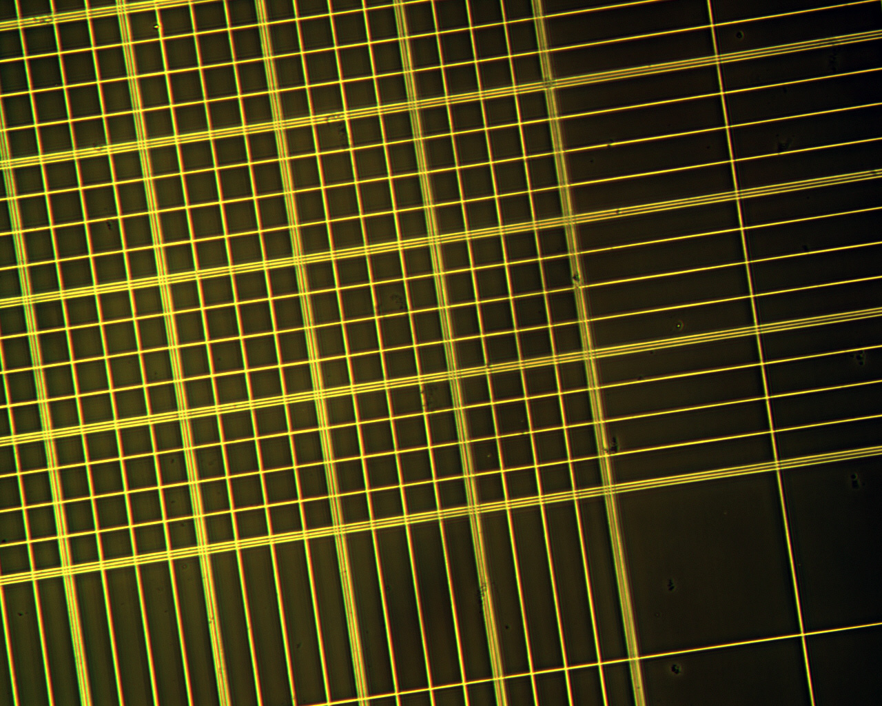
 Empty hemocytometer grid at 100x power.
- The original suspension must be mixed thoroughly before taking a sample. This ensures the sample is representative, and not just an artifact of the particular region of the original mixture it was drawn from.
- An appropriate dilution of the mixture with regard to the number of cells to be counted should be used. If the sample is not diluted enough, the cells will be too crowded and difficult to count. If it is too dilute, the sample size will not be enough to make strong inferences about the concentration in the original mixture.
- By performing a redundant test on a second chamber, the results can be compared. If they differ greatly, the method of taking the sample may be unreliable (e.g., the original mixture is not mixed thoroughly).
References
- ^ Strober W (2001). "Monitoring cell growth". In Coligan JE, Bierer BE, Margulies DH, Sherach EM, Strober W. Current Protocols in Immunology. 5. USA: John Wiley & Sons. p. A.2A.1. doi:10.1002/0471142735.ima03as21.
Categories:


Wikimedia Foundation. 2010.